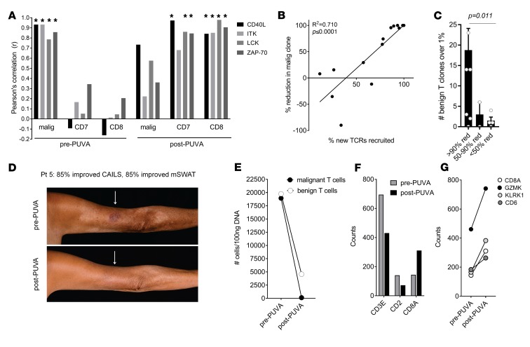
Figure 8. Clearance of the malignant T cell clone after PUVA is associated with recruitment of new CD8+ T cell clones that express markers of antigen-specific activation, are locally expanded, and may be tumor specific.
(A) Markers of TCR-dependent, antigen-specific activation (CD40L, ITK, LCK, and ZAP-70) are strongly associated with malignant T cells before but not after therapy and are strongly associated with benign and CD8+ T cells after, but not before, therapy. Expression of these genes was correlated with malignant T cell number (assessed by HTS), benign T cells (CD7 gene), and CD8+ T cells (CD8A gene) before and after therapy. *P < 0.05. (B) The recruitment of new T cell receptors into the tumor is strongly associated with clearance of the malignant T cell clone. The percentage of total T cell clones bearing antigen receptors not seen before therapy is shown on the x axis and the percentage reduction in the malignant T cell clone is shown on the y axis. A Pearson’s correlation coefficient with a 2-tailed P value is reported. (C) Patients who eradicated or greatly reduced malignant T cells had expanded clonal populations of benign T cells after therapy. The number of expanded benign T cell clones making up greater than 1% of total benign T cells after therapy are shown for patients who had a greater than 90% reduction (>90% red), a 50%–90% reduction (50-90% red), or less than a 50% reduction (<50% red) in malignant T cells after PUVA therapy. The mean and SEM are shown; a Kruskal-Wallis 1-way analysis of variance with a Bonferroni-Dunn post hoc test for multiple means testing was used (α = 0.05). The P value is adjusted for multiple comparison testing. (D–F) An illustrative patient (Pt 5) is shown who had marked reductions in clinical inflammation (D), malignant and total benign T cell numbers in skin (E), but an increase in CD8+ T cell–associated genes (F and G). Malignant and benign T cell numbers were determined by HTS (E) and expression counts for T cell–associated (CD3 and CD2) and CD8-associated (CD8A, GZMK, KLRK1, and CD6) genes (F and G) were measured by NanoString.

