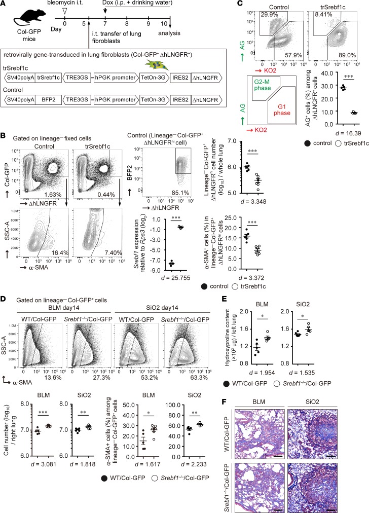
Figure 4. Effect of sterol regulatory element–binding protein 1 on lung fibroblast proliferation and pulmonary fibrosis pathology.
(A) Experimental scheme of the intratracheal transfer of genetically modified lung fibroblasts. (B) Role of active form of Srebf1c (trSrebf1c) in activated fibroblasts in bleomycin-injured lung. Donor fibroblasts were identified by flow cytometry. Graphs show the mean ± SEM (n = 6). A representative result of 2 independent experiments is shown. ***P < 0.001 (2-tailed unpaired Student’s t test). ΔhLNGFRhi gate was defined by induction rates of BFP2 ≥ 80% in all of the control donor fibroblasts. (C) Effect of trSrebf1c on lung fibroblast proliferation in vitro. Cell cycle of genetically modified lung fibroblasts was analyzed by the fluorescence ubiquitination cell cycle indicator. Graphs show the mean ± SEM (n = 5). A representative result of 2 independent experiments is shown. ***P < 0.001 (2-tailed unpaired Student’s t test). (D–F) Lung fibroblast activation and pulmonary fibrosis pathology in Srebf1–/–Col-GFP mice. (D) Number of lung fibroblasts and myofibroblasts in bleomycin- or silica-treated right lungs on day 14. Graphs show the mean ± SEM of n = 5 (BLM WT Col-GFP, SiO2 Srebf1–/–Col-GFP), n = 6 (SiO2 WT Col-GFP), n = 7 (BLM Srebf1–/–Col-GFP). A representative result of 3 independent experiments is shown. *P < 0.05, **P < 0.01, ***P < 0.001 (2-tailed unpaired Student’s t test). (E) Quantification of hydroxyproline content in the whole left lung of bleomycin- or silica-treated Srebf1–/–Col-GFP and WT Col-GFP mice. Graphs show the mean ± SEM of n = 5 (BLM WT Col-GFP, BLM Srebf1–/–Col-GFP, SiO2 Srebf1–/–Col-GFP), n = 6 (SiO2 WT Col-GFP). A representative result of 3 independent experiments is shown. *P < 0.05 (2-tailed unpaired Student’s t test). (F) Masson’s trichrome staining of bleomycin- or silica-treated left lung sections from Srebf1–/–Col-GFP and WT Col-GFP mice at day 14. Scale bars: 100 μm. Representative images of n = 8 (BLM WT Col-GFP, SiO2 WT Col-GFP, SiO2 Srebf1–/–Col-GFP), n = 9 (BLM Srebf1–/–Col-GFP) from 2 independent experiments are shown. (B, C, and D) Effect size (d) is shown on the bottom of the graph. Col-GFP, Col1a2-GFP reporter; ΔhLNGFR, truncated form of human low-affinity nerve growth factor receptor; hPGK, human phosphoglycerate kinase 1; IRES2, internal ribosomal entry site 2; BFP2, mTagBFP2; AG, azami green, KO2, kusabira orange 2; BLM, bleomycin model; SiO2, silica model.

