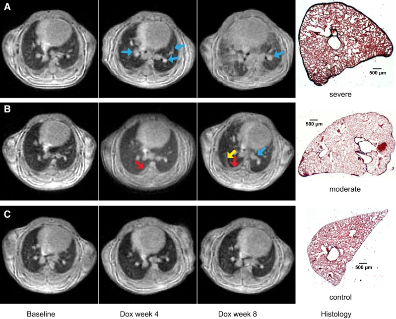
Fig. 4.
Representative magnetic resonance images (repetition time = 6 ms, echo time = 0.108 ms, flip angle = 6.3°) of one doxycycline-fed (Dox) mouse with severe fibrosis (A), one Dox mouse with moderate fibrosis (B), and one control mouse (C) at baseline, Dox week 4, and Dox week 8, plus corresponding histological slides (hematoxylin and eosin stain; original magnification, × 1) at Dox week 8. No pulmonary fibrosis was visually seen at baseline in the Dox cohort and at all 3 time points in the control cohort. Perivascular fibrosis appeared in both Dox mice at Dox week 4 and Dox week 8 (indicated by blue arrows) in A and B. Fibrosis appeared near the pleural region at Dox week 4 (indicated by a red arrow) and extended into the interstitium at Dox week 8 (indicated by a yellow arrow), as seen in B. Apparent interstitial fibrosis also appeared in the Dox mouse in A at Dox week 8. Fibrosis severity varied among different mice as demonstrated in A and B at Dox week 8.

