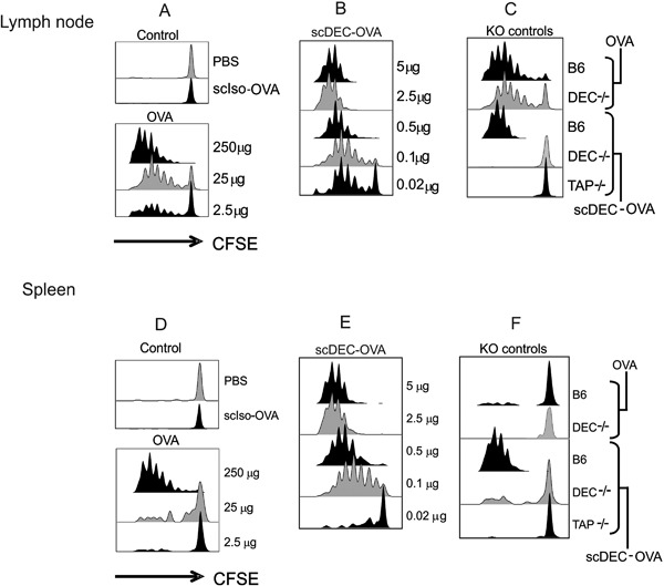
Figure 1.

Antigen presentation to OT‐I T cells following scDEC targeting in vivo. scDEC‐OVA induces stronger in vivo proliferation of OT‐I T cells than OVA alone. C57BL/B6 mice were injected intravenously with 2 × 10⁶ CFSE‐labelled OT‐I T‐cells then graded doses of scDEC‐OVA (A) or OVA (B) subcutaneously 24 h later. Three days after scDEC injection, draining lymph node cells were harvested and the expansion of CD8+ Vα2ß5.1/5.2 evaluated by flow cytometry for CFSE dilution. scDEC‐OVA elicited better presentation of OVA derived peptide than the scCon‐OVA and soluble OVA. In contrast to C57BL/B6 (B6) presentation of peptide from scDEC‐OVA could not be seen in DEC205 K (DEC−/−) and TAP knock out (TAP−/−) mice. On the contrary OVA peptide from B6 and DEC−/− could be presented (C). In (D) to (F) similar data is shown for the spleen. Here as shown in the KO controls presentation was less efficient in the spleen (F). The experiment was repeated several times with similar results.
