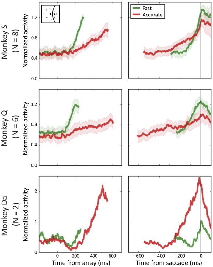
Fig. 5.

Averaged, normalized discharge rates of frontal eye field movement-related neuron samples aligned to array presentation (left) and saccade initiation (right) when the target appears in the movement field on correct fast (green) and correct accurate (red) trials for each monkey. Shaded areas represent means ± SE. For neurons recorded in monkeys Q and S, the average activation at response time was lower on average for accurate relative to fast trials. The opposite was observed for neurons recorded in monkey Da.
