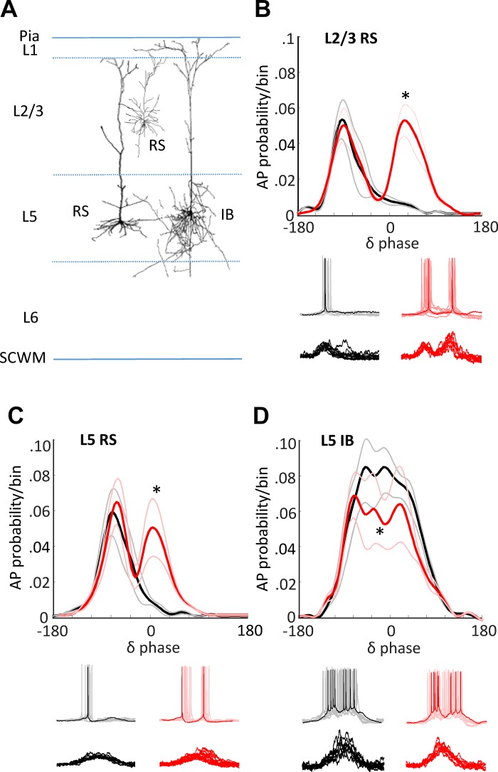
Fig. 6.
Biological model of concurrent theta/delta activity reproduces magnetoencephalography (MEG) outcome-specific dynamics via altered cholinergic excitation. A: schematic of cells targeted in the in vitro parietal cortex slice. Regularly spiking (RS) neurons were recorded in layers 2 and 3 and layer 5. Intrinsically bursting (IB) neurons were recorded in layer 5. Spontaneous delta activity was recorded in the presence of cholinergic agonist (carbachol) at two different concentrations (2 μM, black lines, 6 μM, red lines). B: means ± SE probability of action potential generation in layers 2 and 3 RS neurons binned according to phase of layer 5 field potential delta rhythm. *Significantly larger action potential probabilities (P < 0.05; n = 10 replicates per n = 5 slices/neurons) were seen for the higher level of cholinergic excitation from 30° to 90°. Example traces show activity at resting membrane potential (spikes, upper examples) and at −70 mV [excitatory postsynaptic potentials (EPSPs), lower examples]. C: similar profile of action potential output changes was seen when comparing outputs from layer 5 RS neurons. D: action potential bursts dominated layer 5 IB neuron activity. Elevated cholinergic excitation significantly reduced these burst outputs (*P < 0.05; n = 10 replicates per n = 5 slices/neurons).

