
Figure 2.
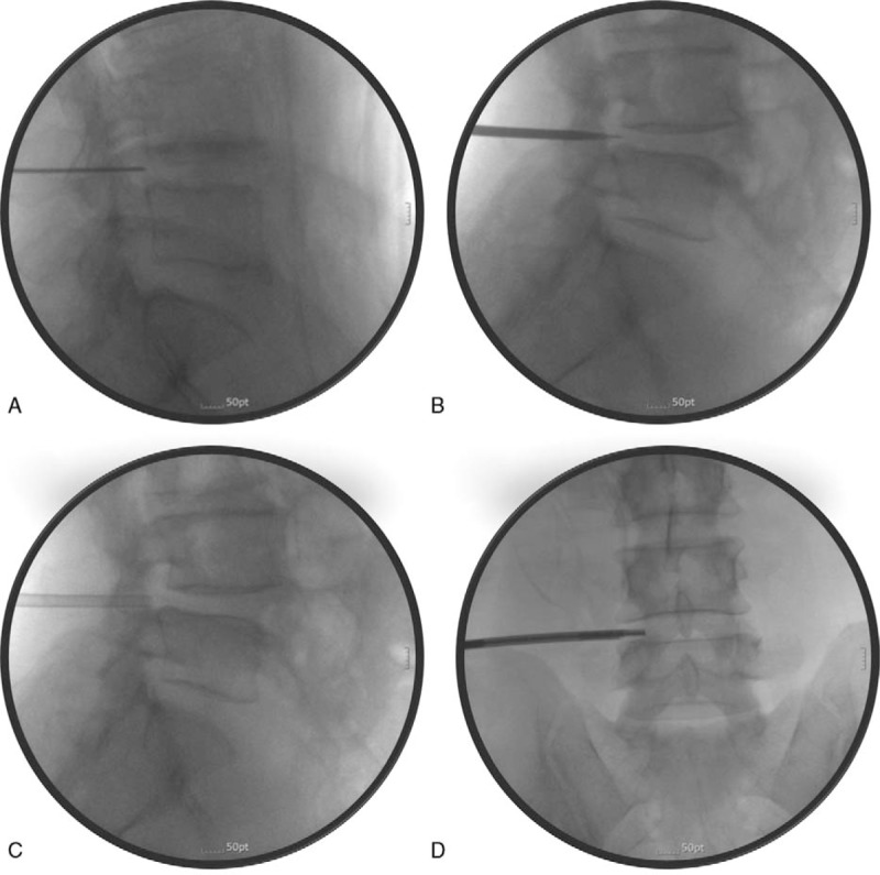
Fluoroscopic images obtained during the percutaneous lumbar foraminoplasty procedure with the Claudicare device. (A) The guidewire was advanced approximately 0.5 cm anteriorly into the target epidural foramen, in the lateral fluoroscopic view. (B) A dilator was inserted via the guidewire into the target foramen until it touched the anterior border of the superior articular process (SAP). (C) After removing the guidewire, a working cannula was inserted via the dilator. The final position of the working cannula was the anterior border of the hypertrophied SAP. (D) A Claudicare drill was inserted via the working cannula and partial removal of the hypertrophied capsule of the SAP commenced from the lateral to medial direction in the AP fluoroscope image.