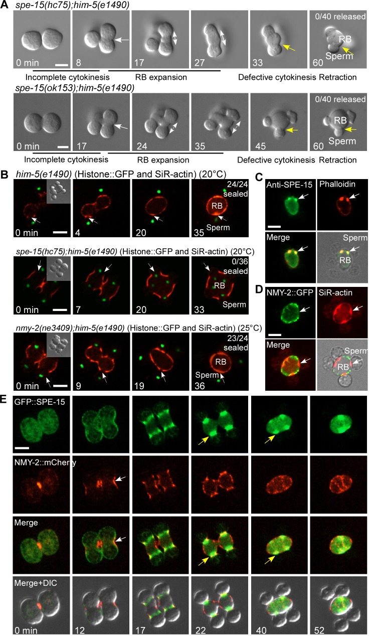
Fig 4. Spermatid release is achieved through myosin VI–mediated cytokinesis.
(A) Time-lapse analysis of meiosis and differentiation in 2 connected secondary spermatocytes dissected from spe-15(hc75);him-5(e1490) and spe-15(ok153);him-5(e1490) males. White arrows indicate the pseudo-cleavage furrow between 2 undifferentiated spermatids. Double-headed arrows indicate RB expansion during spermatid differentiation. Yellow arrows point to the spermatid–RB boundary and indicate defective cytokinesis. Numbers of released spermatids/total spermatids were quantified and are shown in the top right corner. (B) Time-lapse analysis of spermatids and RBs that undergo cytokinesis in the indicated strains expressing Histone::GFP and stained by SiR-actin. White arrows indicate sites of cytokinesis. Numbers of sealed spermatids/total spermatids were quantified and are shown in the top right corner. DIC images of spermatids and RBs at the start time point (0 min, insets) are shown. (C and D) Light and fluorescence images of an RB with 4 attached spermatids stained by anti-SPE-15 antibody and phalloidin (C) or expressing NMY-2::GFP and stained by SiR-actin (D). Arrows point to the connection between the spermatid and RB. (E) Time-lapse images of meiosis and differentiation in 2 connected secondary spermatocytes dissected from him-5 males expressing GFP::SPE-15 and NMY-2::mCherry. White arrows indicate the pseudo-cleavage furrow with enriched NMY-2. Yellow arrows indicate accumulation of SPE-15 at the cleavage furrow between spermatids and RB. Scale bars: 5 μm. DIC, differential interference contrast; GFP, green fluorescent protein; NMY-2, non-muscle myosin 2; RB, residual body; SiR, silicon-rhodamine; SPE-15, defective spermatogenesis 15.

