
Fig. 4-E.
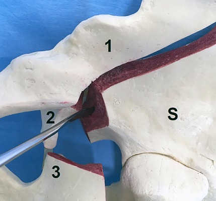
Photograph with a medial view of a Sawbones fracture model (Pacific Research Laboratories) of a right hemipelvis showing a dislocated anterior column posterior hemitransverse acetabular fracture with the displaced anterior column (1), quadrilateral plate (2), posterior hemitransverse fracture (3), and the stable part (S) of the right hemipelvis. The raspatory is used for subchondral disimpaction of an acetabular dome fragment directly through the fracture with the femoral head under traction.