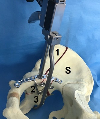
Fig. 4-K.

Photograph of the medial view of a Sawbones fracture model of a right hemipelvis depicting a dislocated anterior column posterior hemitransverse acetabular fracture with the displaced anterior column (1), quadrilateral plate (2), posterior hemitransverse fracture (3), and the stable part (S) of the right hemipelvis. A collinear reduction clamp can be used for compression of the posterior column to the anterior column.
