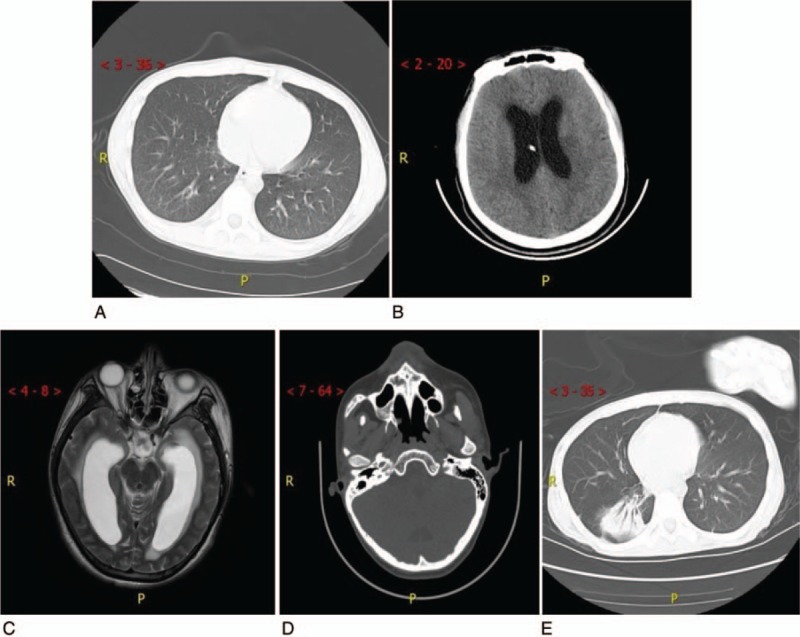
Figure 1.

Computed tomography (CT) image after admission. (A) Nonenhanced pulmonary CT on Feb. 2, 2018. (B) Nonenhanced skull CT scan shows intracranial drainage with increased reduction of intracranial pressure. (C) Skull MRI upon first CSF culture shows XDRAB infection. (D) Nonenhanced skull CT scan after transfer to the rehabilitation unit. (E) Nonenhanced pulmonary CT scan after transfer to the rehabilitation unit. CSF = cerebrospinal fluid, CT = computed tomography, MRI = magnetic resonance imaging, XDRAB = extensively drug-resistant Acinetobacter baumannii.
