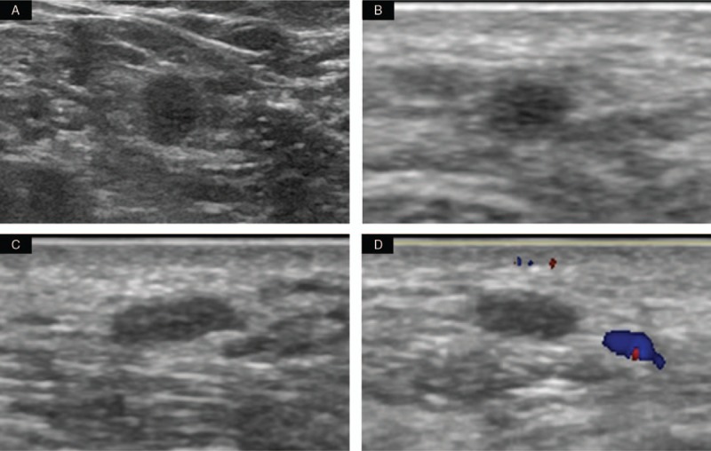
Figure 1.

Ultrasound findings of a 78-year-old woman. (A) Grayscale sonogram of the left supraclavicular area demonstrated a lymph node with abnormal structure, which was approximately 6 × 6 mm in size. (B–D) Two oval, well-circumscribed, parallel nodules are shown in the upper-outer quadrant near the mastectomy scar. (B) Grayscale sonogram of the first nodule shows that it is approximately 4 × 2 × 3 mm in size. (C–D) Grayscale sonogram and color Doppler flow imaging of the second nodule show that it is approximately 6 × 2 × 4 mm in size.
