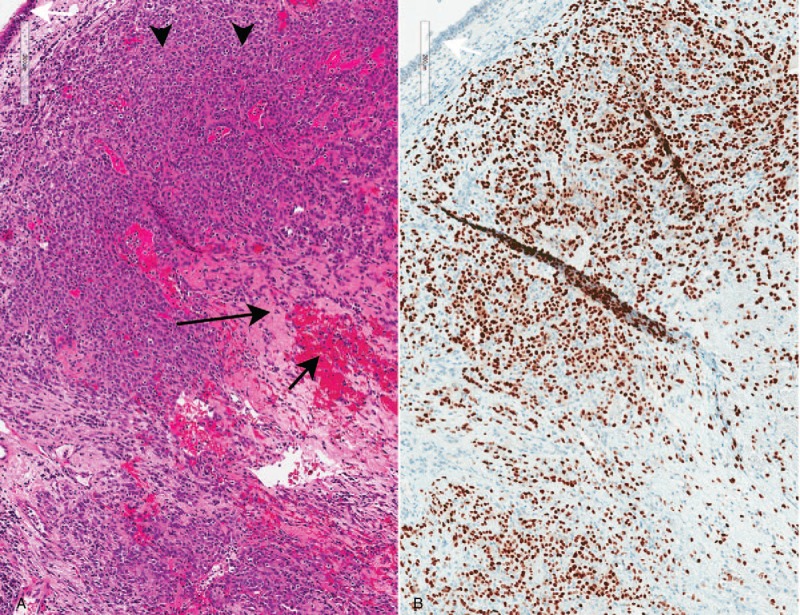
Figure 3.

Pathologic findings: sclerosing pneumocytoma in an endobronchial biopsy. A. Sheets of cytologically bland neoplastic cells fill the bronchial mucosa (arrowheads, hematoxylin-eosin, 10×). Sclerotic (long black arrow) and hemorrhagic (short black arrow) areas are appreciable, even in this small sample. White arrow: respiratory epithelium. B. The neoplastic cells are positive for TTF-1 (10×). White arrow: respiratory epithelium.
