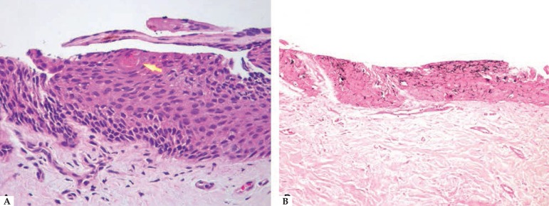
Figure 3.
Histopathological analysis revealed: A - Hematoxylin & eosin (x200) moderate acanthosis of the nail matrix, with proliferation of matrical onychocytes with small monomorphic nuclei with condensed chromatin and scant cytoplasm. Arrow: area of keratinization simu lating a squamous eddy. B - Fontana Masson (x100): melanin within the cytoplasm of matrical onychocytes and sparse melanocytes in the dendrites

