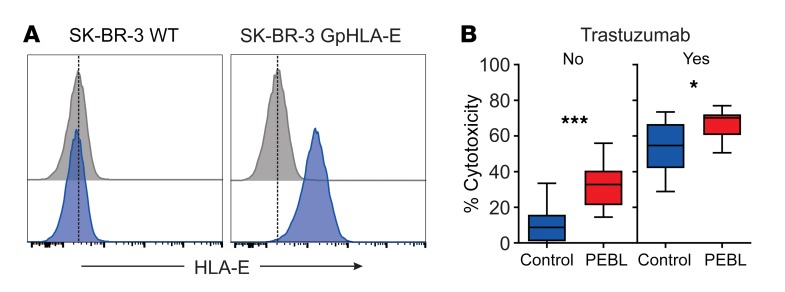
Figure 5. Downregulation of NKG2A increases ADCC activity.
(A) Expression of HLA-E in the SK-BR-3 cell line transduced with HLA-E plus HLA-G signal peptide (GpHLA-E) or nontransduced (WT). Cells were labeled with APC-conjugated anti–HLA-E APC (blue) or isotype-matched nonreactive antibody (gray). (B) Four-hour cytotoxicity with NK cells transduced with anti-NKG2A PEBL or GFP alone (Control) against SK-BR-3-GpHLA-E cells expressing luciferase. BrightGlo was added after 4 hours of coculture, and luminescence was measured using a Flx 800 plate reader. Box (25th–75th percentile, median) and whiskers (minimum-maximum) graphs indicate the collective results of triplicate measurements obtained with NK cells from 2 donors, at a 1:1 or 1:2 E/T. Trastuzumab was added at 10 μg/ml. Horizontal bars correspond to median value. ***P = 0.0001; *P = 0.018, t test.

