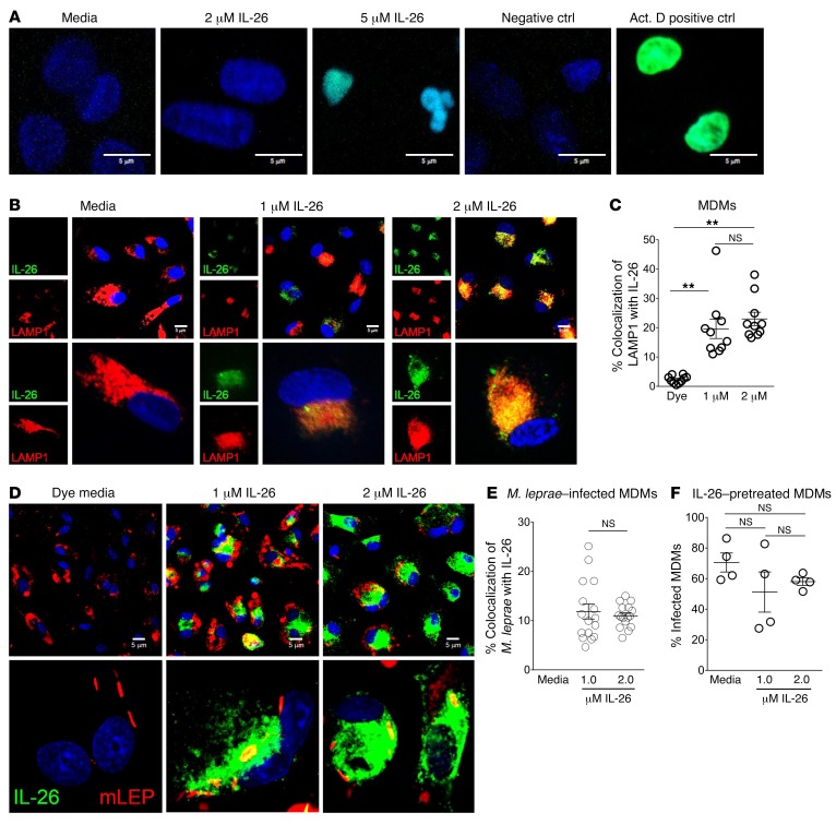
Figure 6. IL-26 is taken up by MDMs and colocalizes with M. leprae.
(A) Human MDMs were treated with IL-26 overnight. Cells were washed and fixed, and apoptosis was determined using a TUNEL (green) assay. Nuclei were stained with DAPI (blue). Data shown are from 5 individual donors. Scale bars: 5 μm. (B) Human MDMs were treated with Alexa 488–IL-26 (green) overnight. Cells were washed, fixed, and immunolabeled with anti-LAMP1 Ab (red). Nuclei were stained with DAPI (blue). Data shown are representative of 5 individual donors. Scale bars: 5 μm. Bottom row magnification, ×630. (C) Colocalization of LAMP1 (red) and IL-26 (green) was quantified with ImageJ. Data represent the mean percentage of colocalization ± SEM (n ≥ 50 cells from 3 donors). **P < 0.01, by repeated-measures 1-way ANOVA. (D) Human MDMs were treated with Alexa 488–IL-26 (green) for 30 minutes and infected with M. leprae (red) overnight. Cells were washed and fixed. Nuclei were stained with DAPI (blue). Media contained Alexa Fluor 488 dye as a control. Data shown are from 4 individual donors. Scale bars: 5 μm. Bottom row magnification, ×630. (E) Colocalization of M. leprae (red) and IL-26 (green) was quantified with ImageJ. Data represent the mean percentage of colocalization ± SEM (n ≥ 40 cells from 4 donors). (F) Quantification of M. leprae–infected MDMs following 30 minutes of treatment with IL-26. Data represent the mean percentage ± SEM (n = 4).

