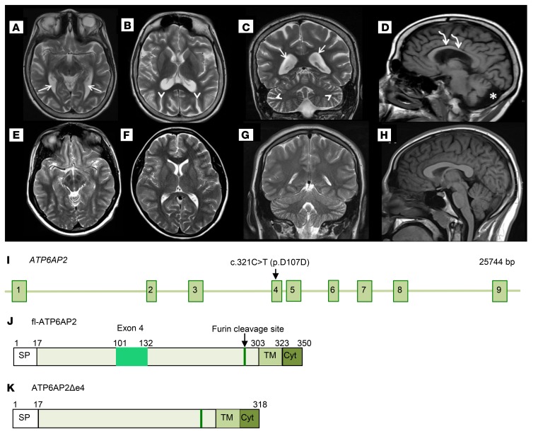
Figure 2. Cerebral atrophy in a patient carrying the ATP6AP2 c.321C>T (p.D107D) variant.
(A–H) Brain MRI of the 15-year-old male patient carrying the c.321C>T variant in exon 4 (OMIM 300423) and of an age-matched normal subject. Patient axial T2-weighted MR brain images (A and B) and normal subject brain images (E and F) for comparison. Patient coronal T2-weighted MR image (C) and normal subject (G). Patient MR images (A–C) exhibit diffuse parenchymal volume loss of gray and white matter. This is observed in the white matter tracts that travel adjacent to the prominently enlarged lateral ventricles (white arrows), as well as the prominent sulci (white arrowheads) in the cerebral hemispheres and cerebellum. Supratentorial and infratentorial parenchymal volume loss involves the cerebral hemispheres and cerebellum symmetrically. Sagittal T1-weighted MR brain image (D) compared with normal subject (H) demonstrates significantly reduced white matter volume of the corpus callosum (curved white arrows). There is also significant white matter volume loss in the patient cerebellum (D) with increased cerebrospinal fluid (asterisk) in the prominent posterior fossa mega cisterna magna. (I) ATP6AP2 genomic locus and positions of variant c.321C>T (p.D107D). (J and K) Schematics of ATP6AP2 and ATP6AP2Δe4.

