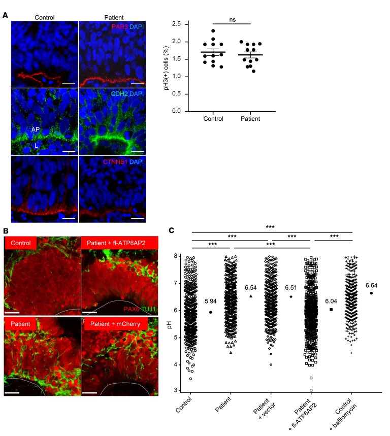
Figure 6. Abnormal “in vitro corticogenesis” of patient iPSC-derived neurons.
(A) Left: Laminar organization of proliferative and differentiation compartments of “cortical” rosettes derived from control and patient iPSCs after 20 days in vitro (DIV). Normal expression pattern of apical marker PAR3 and adherens junctions markers CDH2 and CTNNB1. AP, apical pole; L, lumen. Scale bars: 10 μm. Right: Phospho–histone 3 (pH3) immunostaining did not reveal differences in progenitor proliferation between control and patient. Data show individual values and mean ± SEM; n = 12 per group. P = 0.341 (ns), Student’s t test (unpaired, 2-tailed). (B) Immunostaining with PAX6 and TUJ1 shows interspersed neurons in the progenitor zone of patient-derived rosettes. This phenotype is rescued by re-expression of fl-ATP6AP2. Lines: luminal surface. Scale bars: 25 μm. (C) Significantly increased median pH in patient cells in early differentiating neurons. Data show individual values and median; n = 463–550 measurements from 5 independent experiments. ***P < 0.001, Kruskal-Wallis test (χ2 = 245.97, df = 4, P < 0.001) followed by Dunn’s multiple-comparisons test.

