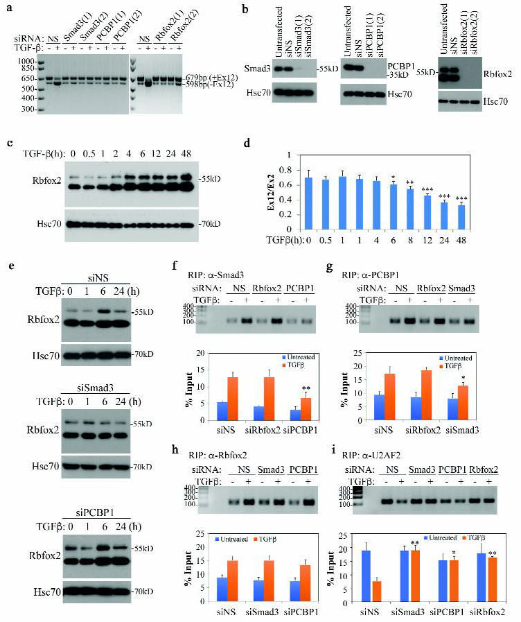
Fig. 2. TGF-β-mediated TAK1 exon 12 exclusion requires Smad3, PCBP1 and Rbfox2.

(a) RT-PCR detection of TAK1 isoforms in NMuMG cells transfected with siRNA and treated with TGF-β for 72 h. NS indicates Non-specific siRNA.
(b) Western blot showing Smad3, PCBP1 and Rbfox2 knockdown in NMuMG cells.
(c) Western blot showing Rbfox2 expression is induced by TGF-β in NMuMG cells.
(d) qRT-PCR analysis of TAK1 exon 12 ratio to TAK1 standard exon 2 in NMuMG cells treated with TGF-β at various time points. Data are shown as mean ± SD (n=3), statistically significant difference between treated and untreated samples is indicated, *p<0.05, **p<0.01, *** p<0.001.
(e) Western blot showing that Smad3 but not PCBP1 is required for TGF-β-induced Rbfox2 expression in NMuMG cells.
(f-i) Recruiting Smad3, PCBP1, Rbfox2 and U2AF2 to TAK1 pre-RNA by TGF-β. Top panels: standard PCR and gel analysis of RIP samples with a TAK1 pre-mRNA primer set, TGF-β treatment was for 2 h; bottom panel: qRT-PCR analysis of samples used in top panels. Data are shown as mean ± SD (n=3), statistically significant difference comparing siRNA transfected to control siNS transfected TGF-β-treated sample is indicated, *p<0.05, **p<0.01.
