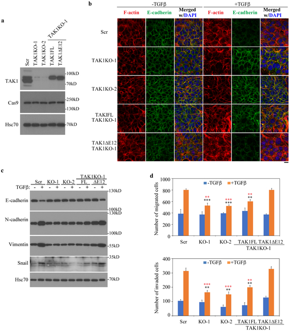
Fig. 6. TAK1∆E12 induces EMT and invasion responses by TGF-β in breast cancer Met-1 cells.
(a) Generation of Met-1 cells with TAK1 deletion by Crispr/Cas9 and reconstitution of Met-1 cells that stably expressing TAK1FL and TAK1∆E12. Two clones that with sgRNA targeting either exon 2 (Tak1 KO-2) or Exon 5 (TAK1 KO-1) were selected, then TAK1FL or TAK1∆E12 was stably expressed in TAK1 KO-1 Met-1 cells. Scr indicates the cells that were transfected with scramble sgRNA.
(b) TAK1∆E12 promotes EMT responses in Met-1 cells. Cells were treated with TGF-β for 3 days, then immunostained for F-actin (phalloidin) and E-cadherin. Bar = 20 μm.
(c) Western blot analyses of expression of the epithelial marker E-cadherin and mesenchymal marker N-cadherin, Vimentin and Snail in Met-1 cells carrying various TAK1 vectors after TGF-β treatment for 3 days.
(d) TAK1∆E12 is required for TGF-β-mediated migration and invasion. Quantitation of migration and invasion were performed by counting cells that across membrane. Data are represented as mean ± S.D. (n=3), statistically significant difference comparing to TGF-β-treated Scr cells or TAK1∆E12-expressing TAK1 KO-1 cells is indicated using black or red asterisks respectively. **p<0.01, ***p<0.001.

