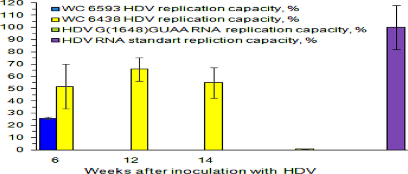
Fig. 6. Replication capacity of intrahepatic HDV RNA genomes harvested from woodchucks M6593 and F6438.

Y axis represents replication capacity of HDV genomes, %. The values of the replication capacity (%) are shown as colored bars. Replication capacity is a normalized parameter, which shows how many HDV genomes/cell were produced as a result of transfection per every HDV genome that was used per cell during the transfection procedure. The value of the replication capacity obtained for in vitro made gel-purified unit length HDV G RNA was used as 100% value. Intrahepatic RNA was isolated from the liver tissue samples obtained from F6438 at weeks +6, +12, and +14 and analyzed. Due to very low intrahepatic HDV G RNA content, the amounts of HDV G RNA that are sufficient for transfection could only be isolated for the week +6 sample of M6593. The time after HDV inoculation (week) is shown at the bottom. As a negative control, the in vitro made gel-purified HDV RNA bearing the mutation G(1648)GUAA, which replication capacity was expected to be profoundly reduced (59), was used during the transfection.
