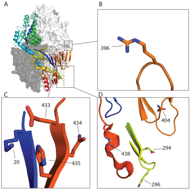
Figure 3: HMPV residues identified thus far involved in low pH mediated fusion.
A) The pre-fusion homo-trimeric structure of HMPV F of NL/1/00 (pdb: 5WB0) with residue positions identified for low pH fusion highlighted including (B) 396 (C) 20, 433, 434, 435(D) 294, 296, 396, 404 and 438. The HRB region was predicted using LearnCoil score for viral membrane fusion proteins as described (39, 40) and stretches from position 453–487 of the fusion protein.

