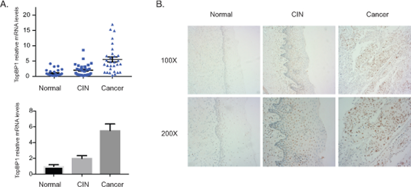
Figure 1.

Increased levels of TopBP1 in HPV-positive cervical cancer tissues. A). RT-PCR analysis of TopBP1 mRNA levels in 29 normal tissues (Normal), 31 cervical intraepithelial neoplasias (CIN), and 30 cervical cancer (CA). The TopBP1 level was shown as as 2-ΔΔCT in biopsies of cancer tissues versus normal tissues. GAPDH was used as internal control and for normalization of the data. The results were shown in both scatter plot figure format and bar figure format. B). Representative immunohistochemical staining of TopBP1 in 10 normal tissues, 10 cervical intraepithelial neoplasias, and 10 cervical cancers.
