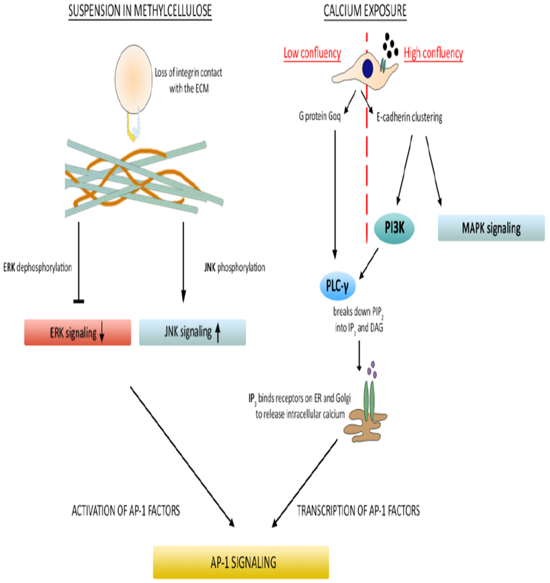
Fig 3. Diagram of intracellular signaling pathways activated by differentiation stimuli.

A representation of the intracellular signaling pathways known to be activated by differentiation stimuli used in Figures 1 and 2. Following suspension in methylcellulose, phosphorylation and signaling activity of extracellular signal-regulated kinase (ERK) decreases. Phosphorylation and signaling activity of c-Jun N-terminal kinase (JNK) increases. This leads to activation of AP-1 transcription factors and subsequent transcriptional expression of differentiation markers. For cells plated at low confluency, exposure to extracellular calcium activates the G protein Gαq, which in turn activates phospholipase C gamma (PLC-γ). PLC-γ hydrolyzes the second messenger phosphatidylinositol 4,5-bisphosphate (PIP2) and activates a series of reactions that lead to the transcription of AP-1 subunits, AP-1 signaling and subsequent transcriptional expression of differentiation markers. For cells plated at high confluency, exposure to extracellular calcium leads to E-cadherin clustering, activating mitogen-activated protein kinase (MAPK) and phosphoinositide 3-kinase (PI3K) activity. PI3K activates PLC-γ and here, the pathway converges with that of low confluency cells treated with calcium terminating in AP-1 signaling and transcription of differentiation markers.
