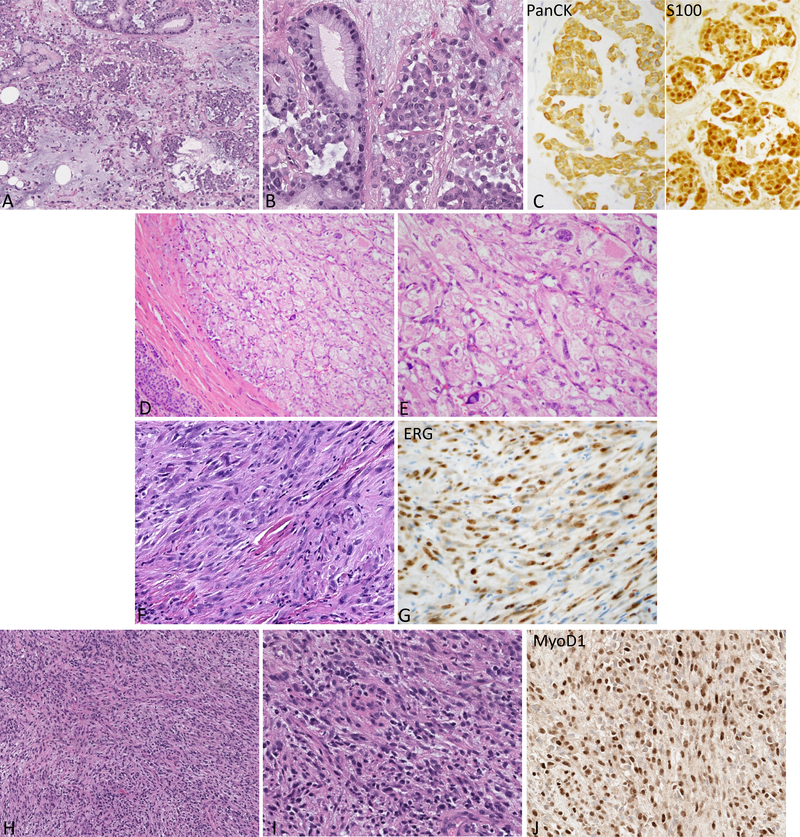
Figure 4.
Representative H&E and immunophenotype of the sarcomas with novel fusions.
A-C: Soft tissue myoepithelial tumor shows epithelioid to plasmacytoid cells in aggregates in a myxoid background with scattered areas showing distinct gland formation. The tumor is strongly and diffusely positive for Pan-CK and S100 by IHC. (A: 200x, B-C: 400x)
D-E: Perivascular epithelioid cell tumor (PEComa) in the pancreas. The tumor is well circumscribed at the periphery and demonstrates large polygonal cells with eosinophilic to clear cytoplasm. (D: 200x, E: 400x)
F-G: Pseudomyogenic hemangioendothelioma composed of spindle cells with moderate amount of eosinophilic cytoplasm and is positive for ERG immunostain. (400x)
H-J: Rhabdomyosarcoma (FUS-TFCP2) arising in the maxillary gingiva demonstrating monomorphic oval to spindle cells in an inflammatory background. The tumor is strongly positive for MyoD1 by IHC. (H: 200x, I-J: 400x)

