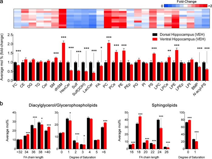
Fig. 2. Distinct lipid composition of dorsal and ventral hippocampus in adult rats.
a LC-MS analysis of dorsal and ventral hippocampus macrodissected from adult rats injected with vehicle (VEH). For lipid nomenclature, see Methods section. Bar graphs indicate fold-change of average relative mol% of all lipids measured, normalized to dorsal hippocampus (mean ± SEM, N = 9 for dorsal hippocampus, N = 10 for ventral hippocampus). Upper panel, heatmap indicates individual plot of normalized average mol% of each lipid class in the ventral hippocampus. Values represented in gradient color, blue indicates below 1 and red indicates above onefold-change, respectively, normalized to dorsal hippocampus. Descriptive statistics listed in Supplementary Table 1. *p < 0.05, **p < 0.01, and ***p < 0.001 in two-tailed Student’s t test. b LC-MS analysis of diacylglycerol/glycerophospholipid and sphingolipid acyl chain. Values expressed as average mol% of total lipid measured, normalized to dorsal hippocampus. Lipids were classified per total acyl carbons and degree of unsaturation (mean ± SEM, N = 9 for dorsal hippocampus, N = 10 for ventral hippocampus). Descriptive statistics listed in Supplementary Table 2. *p < 0.05, **p < 0.01, and ***p < 0.001 in two-tailed Student’s t test

