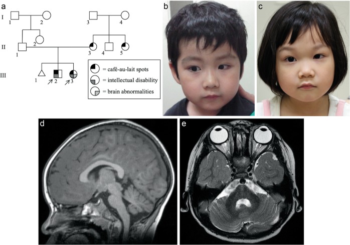
Fig. 1. The family pedigree, photograph, and brain MRI of Patient 1 and 2.
a Three-generation pedigree of the family in this study. Each symbol is annotated below. b, c Photograph of patients 1 and 2. Macrocephaly was noted in patient 1, but microcephaly and bulbous nose were noted in patient 2. d, e Brain MRI of patient 2 (d axial FLAIR image view, e sagittal T1-weighted view). MRI reveals mild hypoplasia of the cerebellum and brainstem. MRI magnetic resonance imaging

