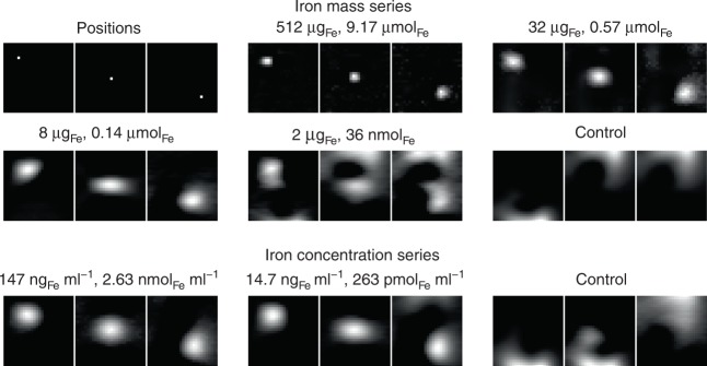
Fig. 2.
Sensitivity of the developed MPI brain scanner. 50 μl samples of Perimag with varying dilutions are moved to three positions on the FOV diagonal (upper 6 images). The sample is considered to be detected if the movement of the sample correlates to the signal shift in the image. Below 2 μgFe this correlation fails. The same procedure is done with an ellipsoid of 134 ml filled with different concentrations (lower 3 images). At a concentration of 14.7 ngFe ml−1 (263 pmol Fe ml−1, 2 μgFe total) the position is still detectable while it fails at concentrations below that value

