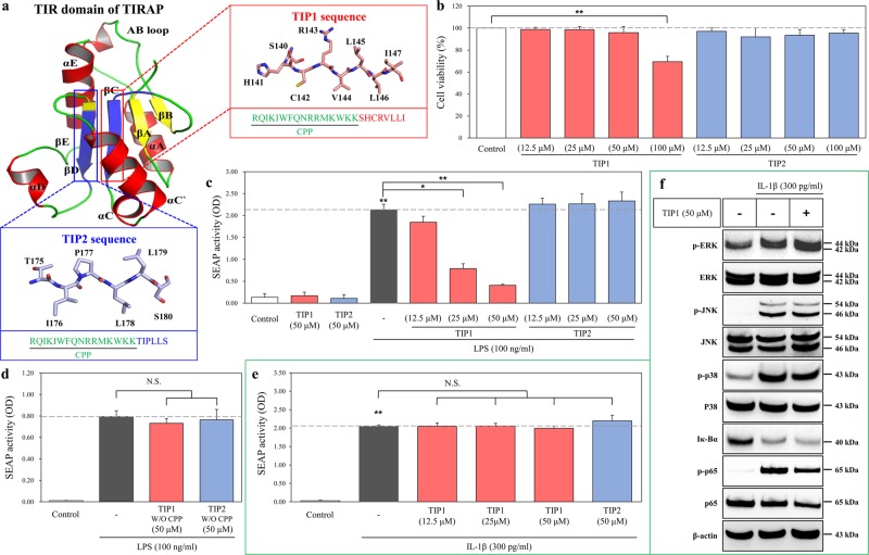
Fig. 1. Design of the TLR decoy peptides derived from the TIR domain of TIRAP.
a TLR-inhibitory peptides (TIP) 1 and 2 were designed using the sheet structures of βC (red box) and βD (blue box), respectively, and then conjugated with cell-penetrating peptide (CPP; green color). b HEK-Blue hTLR4 cells were treated with various concentrations of TIP1 (12.5, 25, 50, or 100 µM) or TIP2 (12.5, 25, 50, or 100 µM) for 24 h. Cell viability was measured on a microplate reader by the MTS assay. c The activation of NF-κB was measured by a SEAP activity assay after the treatment of HEK-Blue hTLR4 cells with the different indicated concentrations of peptides for 1 h in the presence or absence of LPS for 24 h. d The activation of NF-κB was measured by the SEAP activity assay after the treatment of HEK-Blue hTLR4 cells with the TIP1 or -2 peptide not conjugated with CPP: TIP1 or TIP2 without (W/O) CPP. e NF-κB activity was measured by a SEAP activity assay upon the treatment of HEK-Blue IL-1R cells with different concentrations of peptides for 1 h before the treatment with IL-1β (300 pg/ml) for the next 24 h. f HEK-Blue IL-1R cells were treated with TIP1 (50 µM) for 1 h in the presence or absence of IL-1β (300 pg/ml) for 30 min. The amounts of p-ERK, ERK, p-JNK, JNK, p-p38, p38, Iκ-Bα, p-p65, and p65 were measured by western blotting in the total protein extract. β-Actin served as a loading control. The data shown represent at least three independent experiments (n ≥ 3), and bars represent means ± SEM (*P < 0.05, **P < 0.01). N.S.: not significant

