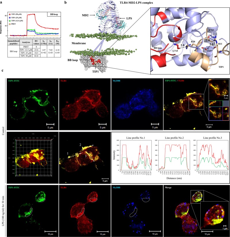
Fig. 4. Characterization of the antagonistic effects of TIP1.
a The potential binding of TIP1 to the BB loop of the TIR domain, as confirmed by SPR analysis. The peptide sequence of the BB loop (710-RDFIPGVAIAA-720) was conjugated with biotin in tandem to facilitate coating of the sensor chip with the peptides. b An illustration of TIP1 binding to the BB loop region of the TLR4-TIR domain. The BB loop is colored red and TIP1 light yellow. The TLR4 extracellular and TM domains are shown as the cylindrical-cartoon model, and the TIR domain is shown as the space-filling model. LPS is colored orange, and phosphate atoms are shown as mauve beads. The hydrogen bond (H-bond)-forming residues are shown as stick models. Digits indicate the distance between H-bond forming atoms in the angstrom unit. c THP1 cells were treated with FITC-conjugated TIP1 for 1 h before treatment with LPS (100 ng/ml) for 30 min. The expression levels of TLR4 and MyD88 and the localization of TIP1-FITC were determined by immunofluorescent staining and confocal microscopy. Orthogonal projections, 3D image reconstruction, and fluorescence intensity profiles of confocal images show localization of TIP1-FITC and TLR4

