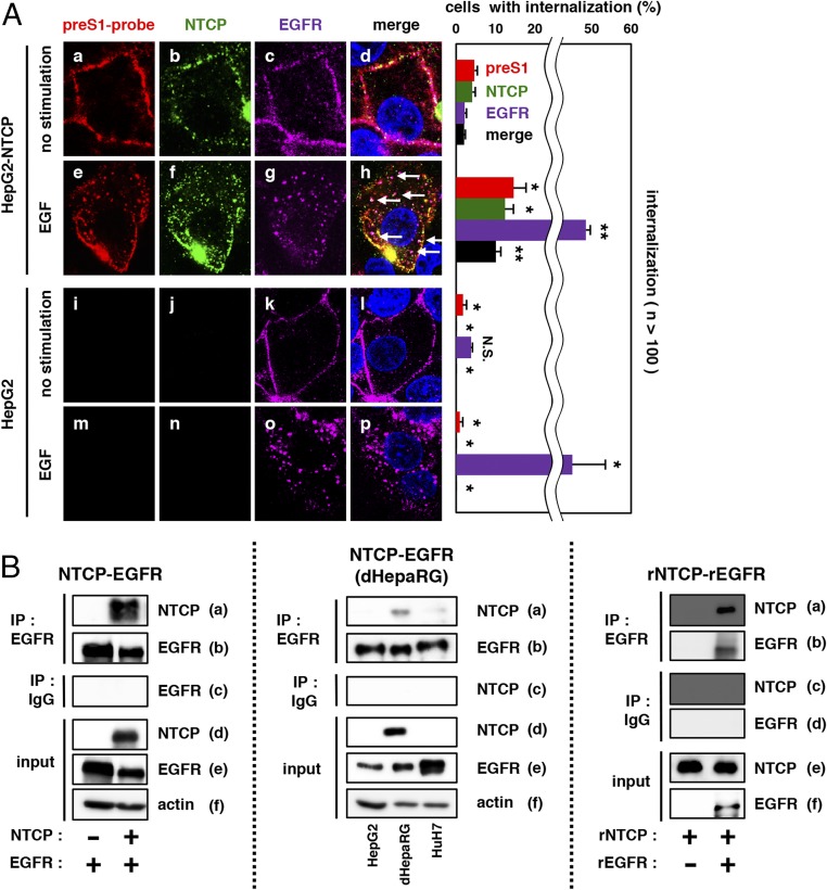
Fig. 3.
EGFR relocalization triggers the preS1–NTCP internalization. (A) HepG2-NTCP or HepG2 cells attached with preS1-probe at 4 °C were stimulated with EGF or left unstimulated (no stimulation) at 37 °C for 30 min and observed by confocal microscopy (preS1-probe, red; NTCP, green; EGFR, purple; nucleus, blue). White arrows indicate the colocalization of preS1, NTCP, and EGFR (Left, d and h). The percentages of cells showing the internalization for preS1, NTCP, and EGFR are indicated in the graph (Right). **P < 0.01; *P < 0.05. N.S., not significant. (B, Left) 293T cells overexpressing EGFR together with or without NTCP were harvested to immunoprecipitate with anti-EGFR antibody (IP: EGFR) or normal mouse IgG as a negative control (IP: IgG) and to detect NTCP or EGFR in the precipitates. NTCP, EGFR, and actin in the total cell lysate were also detected (input). (B, Center) HepG2, dHepaRG, and Huh7 cells were subjected to coimmunoprecipitation assay for endogenous protein interaction. (B, Right) Recombinant NTCP (rNTCP) was incubated with or without recombinant EGFR (rEGRF) in vitro, which was subjected to coimmunoprecipitation analysis as shown above.

