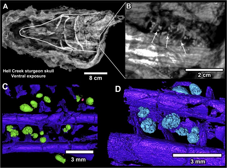
Fig. 6.
Acipenseriform fish with ejecta clustered in the gill region. (A) X-ray of a fossil sturgeon head (outlined, pointing left; FAU.DGS.ND.161.115.T). (B) Magnified image of the X-ray in A showing numerous ejecta spherules clustered within the gill region (arrows). (C and D) Micro-CT images of another fish specimen (paddlefish; FAU.DGS.ND.161.29.T), with microtektites embedded between the gill rakers in the same fashion.

