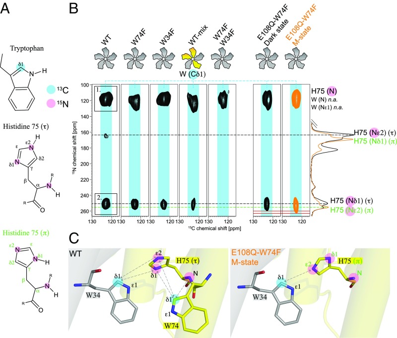
Fig. 4.
Visualizing the cross-protomer H75-W34 contact. (A) For the detection of the cross-protomer H75-W34 contact, (13Cδ1-Trp, 15N3-His)-GPRWT (wild type) and various mutants have been prepared. (B) Two-dimensional TEDOR spectra of GPRWT, GPRW74F, GPRW34F, GPRWT-mix, GPRW74F-W34F, and GPRE108Q-W74F, all with (13Cδ1-Trp, 15N3-His) labeling. The 1D spectra on the Right are from (13C6-15N3-His, 15Nε-Lys)-GPRE108Q (Fig. 3A) in the dark state (black) and M state (orange) and have been plotted to identify the H75(τ) and (π) resonances. Cross peak 1 in GPRWT (Left) is observed in all spectra and corresponds to a mixture of correlations between W34/W74-Cδ1 and H75-N as well as natural abundance correlations between Trp-Cδ1 and Trp-N or Trp-Nε1. Cross peak 2 can be attributed to a correlation between W74 or W34-Cδ1 and H75-Nδ1(τ). Spectra of GPRW74F and GPRW34F confirm that both tryptophan residues contribute to the interaction. The mixed-labeled sample GPRWT-mix consisting of (13Cδ1-Trp)-GPRWT and (15N3-His)-GPRWT proves that a W34-H75 interprotomer contact exists. M state trapping of (13Cδ1-Trp, 15N3-His)-GPRE108Q-W74F shows a stretching of signal 2 toward the Nε2(π) resonance. (C) The GPRWT sample shows close proximity of H75-Nδ1(τ) to W34-Cδ1 and an intraprotomer contact to W74-Cδ1. The GPRE108Q-W74F sample indicates a turn of H75 so that Nε1(π) and W34-Cδ1 occur in close proximity. Data from PDB ID: 4JQ6 (7).

