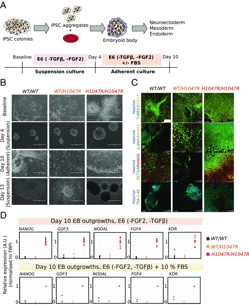
Fig. 4.
Self-sustained stemness in PIK3CAH1047R/H1047R embryoid bodies (EBs). (A) Schematic illustrating the protocol used for EB formation and subsequent adherent culture. E6, Essential 6 medium; FGF2, fibroblast growth factor 2; TGFβ, transforming growth factor β. (B) Representative bright-field micrographs of WT (WT/WT), PIK3CAWT/H1047R (WT/H1047R), and PIK3CAH1047R/H1047R (H1047R/H1047R) cells at baseline (iPSC stage), 4 d (suspension), 10 d (adherent), and 13 d (suspension) following EB formation. PIK3CAH1047R/H1047R iPSC colonies are refractile due to partial dissociation, while stem cell-like colonies emerging from adherent PIK3CAH1047R/H1047R EBs are highly compact. In addition to the floating layers of differentiated cells shown here, WT and PIK3CAWT/H1047R suspension cultures on day 13 also contained larger EB aggregates with complex morphologies and internal differentiation. (Scale bar: 400 μm.) (C) EB outgrowths were fixed on day 10 and stained for TRA-1-60 or costained for TUBB3/SOX17, α-SMA/HAND1 or NANOG/OCT3/4. Hoechst was used for nuclear visualization. Images are representative of two independent experiments, using a single WT clone and two clones of each mutant. (Scale bar: 100 µm.) (D) Real-time quantitative PCR analysis of stemness gene expression in EB outgrowths in E6 medium without TGFβ and FGF2. Individual replicates shown in the panel are from three to four WT clones, two PIK3CAWT/H1047R clones, and four PIK3CAH1047R/H1047R clones (including technical duplicates of the PIK3CAH1047R/H1047R outgrowth cultures). Expression values are in arbitrary units (A.U.). See also SI Appendix, Fig. S5.

