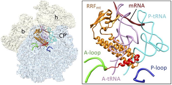
Fig. 2.
Binding positions of bound RRFmt, mRNA, and A- and P-site tRNAs on the mitoribosome. RRFmt binding would be in direct steric clash with the acceptor arm of both aminoacyl- and peptidyl- tRNAs on the large subunit. Coordinates of the mRNA (dark brown) and tRNAs (pink and light blue) were derived from the structure of porcine mitoribosome (25) (PDB ID: 5AJ4). The conserved RRFmt domains and its NTE are colored as in Fig. 1. The NTE of RRFmt lies in close proximity to the functionally important and conserved A (green) and P loops (dark blue). A thumbnail to left depicts an overall orientation of the 55S mitoribosome, with semitransparent 28S (yellow) and 39S (blue) subunits, and overlaid positions of ligands. Landmarks on the thumbnail: h, head, and b, body of the 28S subunit, and CP, central protuberance of the 39S subunit.

