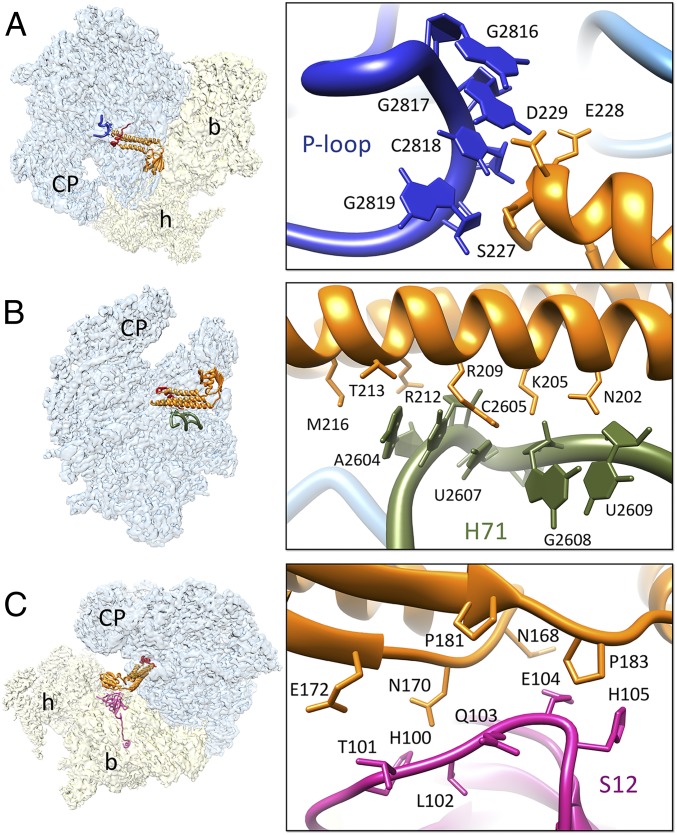
Fig. 3.
Interactions of structurally conserved domains of RRFmt with the 55S mitoribosome. (A) Contacts between the domain I (orange) and the P loop, the 16S rRNA H80 (blue). (B) Interactions between the domain I and H71 (olive green) of the 16S rRNA. (C) Contacts between the ribosomal protein S12 (magenta) and RRFmt domain II. Thumbnails to left depict overall orientations of the 55S mitoribosome, with semitransparent 28S (yellow) and 39S (blue) subunits, and overlaid positions of RRFmt. Landmarks on the thumbnails are same as in Fig. 2.

