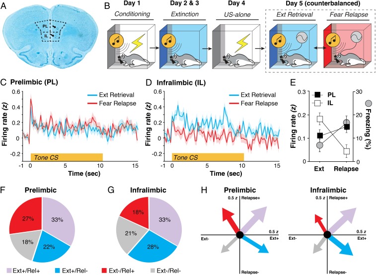
Fig. 1.
Extinction retrieval and fear relapse bidirectionally engage mPFC signaling. (A) Representative histology of electrode placements in PL and IL. (B) Schematized behavioral design. (C and D) CS-evoked firing from PL (C) and IL (D) neurons in retrieval and renewal. (E) Percentage of freezing (gray circles, mean ± SEM) across tests; freezing is overlaid with the 10-s summary of the CS-evoked firing responses. (F and G) Pie charts displaying the proportion of PL and IL neurons in one of four categories based on how neurons responded to presentation of the CS in both retrieval and relapse. (H) Vector plots depicting CS-evoked firing. The tips of the arrows point to the mean CS-evoked responding for a particular quadrant. The thickness of the arrow is proportional to the total number of neurons recorded in either PL or IL. Ext, extinction; Rel, relapse; US, unconditioned stimulus.

