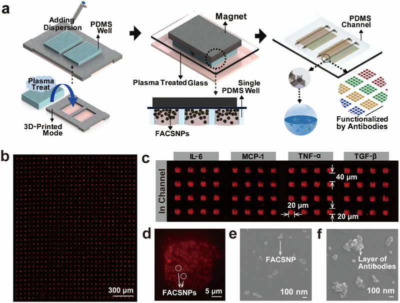
Figure 2. Schematics of the FACSNP microarray fabrication and characterization of the fabricated microarray chip.
(a) Illustrations of the magnet assisted patterning process of the FACSNP microarray. (b) Dark-field microscopy images of the FACSNP microarray on the glass substrate. The FACSNPs confined in PDMS microwells self-assembled into a series of regular square-shape sensing spot arrays with the assistance of the external magnetic field. (c) After the patterning process, the FACSNP microarrays were functioned with four different antibodies in defined sensing areas for multiplex detection of four cytokines. (d) Dark-field microscopy image of individual FACSNPs biosensing spot at higher magnification, showing the well dispersed FACSNPs immobilized in the sensing spot. (e) SEM image of the FACSNPs before antibody function and (f) after successful antibody attachment.

