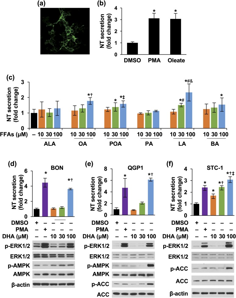
Figure 1.
FFAs stimulate NT secretion from neuroendocrine cells. (a) STC-1 cells were stained with NT antibody and observed by confocal microscope using the 60× oil objective. (b) STC-1 cells were treated with or without PMA (100 nM) or sodium oleate (0.25 mM) for 30 minutes; media were collected and NT EIA performed. *P < 0.05 vs DMSO. (c) BON cells were treated with or without different FFAs at various dosages for 3 hours; media were collected and NT EIA performed. *P < 0.05 vs control; †P < 0.05 vs 10 and 30 μM of OA; ‡P < 0.05 vs 10 μM of POA; #P > 0.05 vs 10 μM of LA; &P > 0.05 vs 30 μM of LA. (d–f) BON, QGP-1, and STC-1 cells were treated with or without different concentrations of DHA or 10 nM PMA (a positive control) for 3 hours; media (upper panels) and cells (lower panels) were analyzed by NT EIA and western blot, respectively. (d) BON cells: *P < 0.05 vs DMSO; †P < 0.05 vs 10, 30 μM DHA; (e) QGP-1 cells: *P < 0.05 vs DMSO; †P < 0.05 vs 10, 30 μM DHA; (f) STC-1 cells: *P < 0.05 vs DMSO; †P < 0.05 vs 10 μM DHA; ‡P < 0.05 vs 30 μM DHA. All data represent mean ± SD. Experiments were repeated at least three times. BA, butyric acid; DMSO, dimethyl sulfoxide; PA, palmitic acid.

