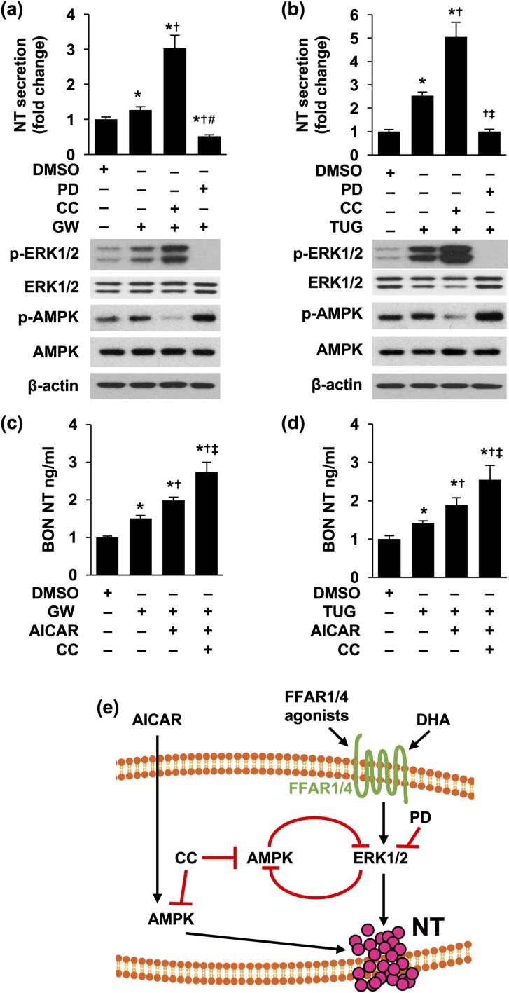
Figure 6.
Inhibition of AMPK signaling further increased NT secretion stimulated by GW 9508 or TUG 891. (a,b) BON cells were pretreated with or without CC (10 μM) for 30 minutes, followed with or without PD (10 nM) for another 30 minutes and then by combined treatment of (a) GW 9508 (10 μM) or (b) TUG 891 (10 μM) with CC or PD for 3 hours. Media were collected and NT EIA performed (upper panels) and cells lysed for western blotting analysis (lower panels). Upper panels: *P < 0.05 vs DMSO; †P < 0.05 vs GW 9508 or TUG 891 alone; ‡P < 0.05 vs GW 9508 or TUG 891 plus CC. (c,d) BON cells were pretreated with CC (10 μM) for 30 minutes and then with or without AICAR (1 mM) for another 30 minutes, followed by combined treatment of (c) GW 9508 (10 μM) or (d) TUG 891 (10 μM) with AICAR or CC for 3 hours. Media were collected and NT EIA performed. *P < 0.05 vs DMSO; †P < 0.05 vs GW 9508 or TUG alone; ‡P < 0.05 vs GW 9508 or TUG 891 plus AICAR. Experiments were repeated at least three times. (e) Summary of signaling downstream of FFAR1 and FFAR4 in regulation of NT release. DHA and FFAR1 or FFAR4 agonists activate FFAR1 and FFAR4 and increase NT secretion through activation of ERK1/2. PD compound inhibits NT secretion by inhibiting MEK/ERK1/2 signaling. Inhibition of MEK/ERK or AMPK releases the inhibitory regulation of each other. DMSO, dimethyl sulfoxide; GW, GW 9508; TUG, TUG 891.

