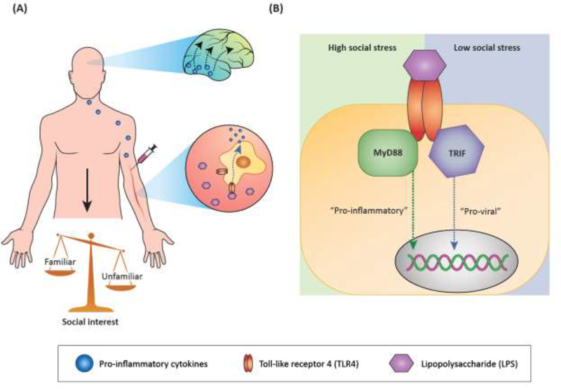
Figure 2. Models of the bidirectional relationships between social behavior and immune function.
(A) Induction of an inflammatory response (and sickness behavior if dose is high enough) with LPS, a structural motif on gram negative bacteria, activates TLR4 on peripheral immune cells, causing release of pro-inflammatory cytokines such as IL-1β and IL-6. Peripheral immune signaling is hypothesized to activate brain regions that can be directly influenced by the periphery, such as brainstem and hypothalamic regions. Through mechanisms that are not yet clear, activated brain regions may induce immune signaling (particularly IL-1β signaling) in brain regions important for motivation, reward, and emotion processing. In turn, social interest shifts from novel/unfamiliar social investigation to increased interaction with familiar social stimuli. (B) Intracellular signaling by TLR4 may be influenced by social context or status. In organisms experiencing high social stress, activation of TLR4 may preferentially induce “pro-inflammatory,” or MyD88-dependent, signaling and subsequent transcriptional signatures. Conversely, in organisms experiencing low social stress, TLR4 activation may preferentially induce “pro-viral,” or TRIF-dependent, signaling and subsequent transcriptional signatures.

