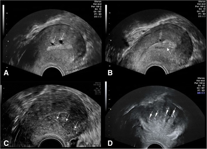
Fig. 1.
Direct and indirect imaging features of adenomyosis on ultrasound. a Small posterior wall myometrial cysts (open arrows). b Poorly defined endo-myometrial interface (solid arrow). c Diffuse myometrial heterogeneity with hyperechoic linear striations (three arrows). d Diffuse asymmetric widening of the posterior myometrial wall with hyperechoic nodules (four arrows)

