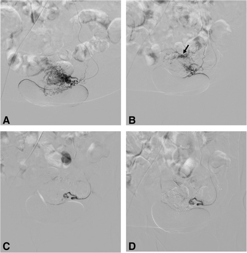
Fig. 3.

Digital subtraction angiography (DSA) images (of the same patient in Fig. 2) with selective injections of the left (a) uterine artery demonstrate with multiple tortuous uterine artery branches and (b) lesion blush (most prominent at the anatomic site of the posterior uterine wall). Right uterine artery injection (not shown) was unremarkable for pathology. Post-embolization DSA images show occlusion of toursous feeding vessels (c) with absence of lesion blush (d)
