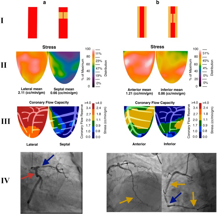
Fig. 7.
Impact of discrete and diffuse disease on relative perfusion images, stress myocardial blood flow (sMBF) and coronary flow capacity (CFC). For illustrative purposes, only two quadrants are shown. aI Normal epicardial vessel and an epicardial vessel with an isolated high-grade discrete stenosis. The normal vessel perfuses the lateral wall and the vessel with discrete stenosis perfuses the septal wall. II sMBF in the lateral wall is near normal (2.11 cm3/min/g), whereas the sMBF in the septal wall is markedly reduced (0.66 cm3/min/g). This wide variability in sMBF has led to an obvious, large severe relative PA in which septal wall uptake is about 30–40% (0.66/2.11 = 0.31) of that in the lateral wall (blue-purple on relative perfusion images). III Coronary flow capacity maps show normal CFC in the lateral wall and a severe reduction in CFC in the septal wall. IV The angiogram in this patient shows that the left circumflex artery, which is a large dominant vessel, is free of disease (red arrow), whereas a visually obvious high-grade stenosis is present in the left anterior descending artery (blue arrow). bI Both vessels are abnormal. They both have diffuse epicardial disease, and one vessel also shows a superimposed high-grade discrete disease. II The impact of diffuse disease is profound. Stress myocardial blood flow is markedly reduced in the anterior wall (1.21 cm3/min/g), which is near the ischemic threshold. The addition of discrete stenosis in the vessel perfusing the inferior wall further reduces sMBF below the ischemic threshold (0.86 cm3/min/g). However, the relative drop in perfusion is mild, as the difference in sMBF between these walls is small. This scenario yields a trivial and nonsignificant relative defect (0.86/1.21 = 71% uptake; yellow zone in the inferobasilar wall). However, CFC maps demonstrate a moderate reduction in flow capacity at the apex (secondary to diffuse disease leading to a base-to-apex gradient) and a severe reduction in flow capacity in the inferior wall due to the combination of discrete and diffuse disease. IV The angiogram in this patient shows that the left anterior descending artery is diffusely diseased and tapers towards the apex (yellow arrow). The right coronary artery has several proximal patent stents with mild in-stent diffuse stenosis, and also tapers distally (yellow arrows). In addition, a high-grade stenosis is seen in the mid right coronary artery (blue arrow)

