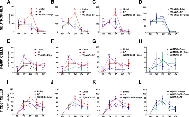
Fig. 4.
Influx of inflammatory cells into wounded tissues: polymorphonuclear neutrophils (a–d), macrophages (e–h), and T cells (i–l). Experimental groups: wounds covered with Linitul (purple line), wounds covered with SF scaffolds (red line), Wharton’s jelly MSCs injected at the wound edge (Wj-MSCs-Edge) (black line), wounds covered with SF scaffolds cellularized with Wharton’s jelly MSCs (Wj-MSCs-SF) (green line), and wounds treated with Wharton’s jelly MSCs injected at the wound edge and cellularized SF scaffolds onto the wound bed (Wj-MSCs-SF+Edge) (blue line). Statistically significant differences (p < 0.05) compared to Linitul (value 1), scaffold (value 2), Wj-MSCs-Edge (value 3), Wj-MSCs-SF (value 4), and Wj-MSCs-SF+Edge (value 5), according to one-way ANOVA

