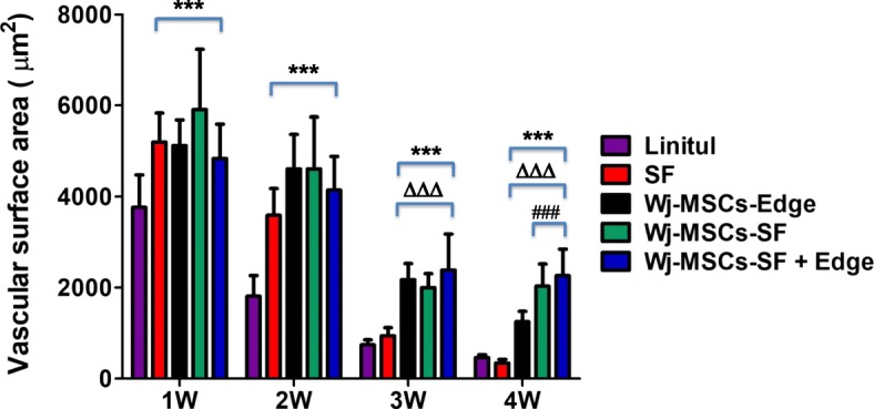
Fig. 5.

Vascular surface area (expressed in square micrometers per field) determined by immunohistochemical analysis of CD31 expression in wound sections. Vascularized area was significantly increased compared to the untreated group (Linitul) (***p < 0.001), SF group (∆∆∆p < 0.001), or Wj-MSCs-Edge-treated group (###p < 0.001), respectively, according to one-way ANOVA. Results are shown as mean ± SD of the most vascularized areas measured in three different mice of each group, corresponding to images made at × 400 magnification. Experimental groups: SF (wounds covered with silk fibroin scaffold), Wj-MSCs-Edge (Wj-MSCs injected at the wound edge), Wj-MSCs-SF (wounds covered with silk fibroin scaffold cellularized with Wj-MSCs), and Wj-MSCs-SF+Edge (wounds treated with Wj-MSCs injected at the wound edge and cellularized silk fibroin scaffold onto the wound bed)
