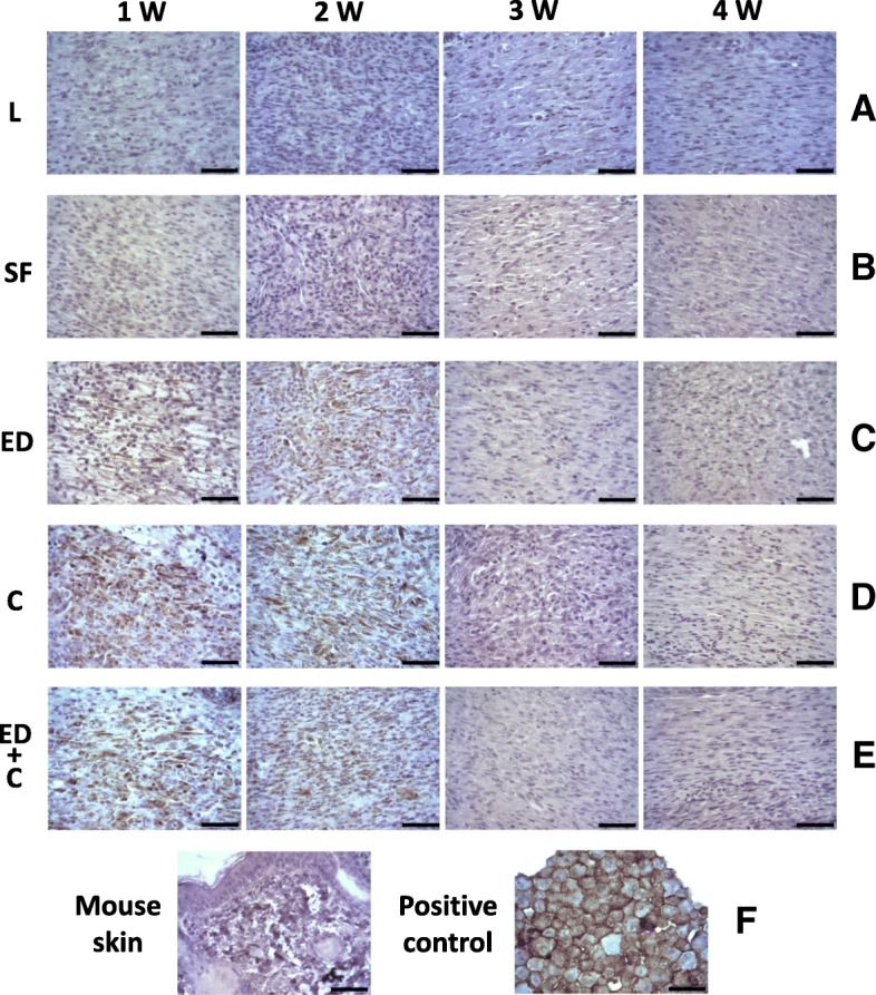
Fig. 7.

a–e Expression of the human mesenchymal stem cell marker CD90 on mouse dermal wounds. Experimental groups: L (wounds covered with Linitul), SF (wounds covered with SF scaffolds), ED (wounds with Wj-MSCs injected at the edge), C (wounds covered with SF patches cellularized with Wj-MSCs), and ED+C (wounds with SF patches cellularized with Wj-MSCs over the wound bed and Wj-MSCs injected at the edge). Immunostaining with the anti-human CD90 antibody of a mouse skin section and a pellet made of Wj-MSCs served as negative and positive controls, respectively (f). Scale bar 50 μm
