Fig. 5.
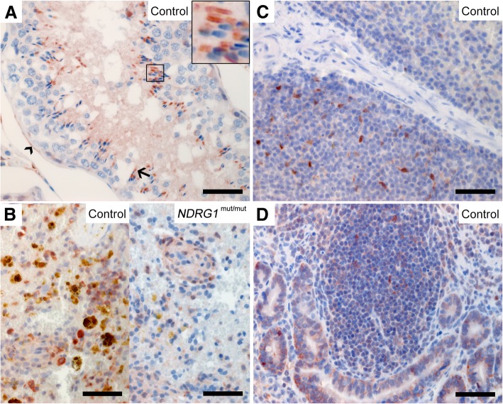
Expression of NDRG1 in developing spermatids and mesenchymal cells. NDRG1 is present in the midpiece (arrow and inset) of the spermatids and the myoepithelial cells (arrowhead) surrounding the seminiferous tubules (a), the ellipsoids and a subpopulation of the leukocytes in the spleen (b), follicles in the spleen (c) and the intestinal Peyer’s patches (d). The NDRG1 localization in the spleen of the NDRG1mut/mut Alaskan malamute is similar to the control (c). Note the extensive hemosiderin deposits (yellow-brown granules) in the spleen of the control dog. Bar 50 μm

