Fig. 1.

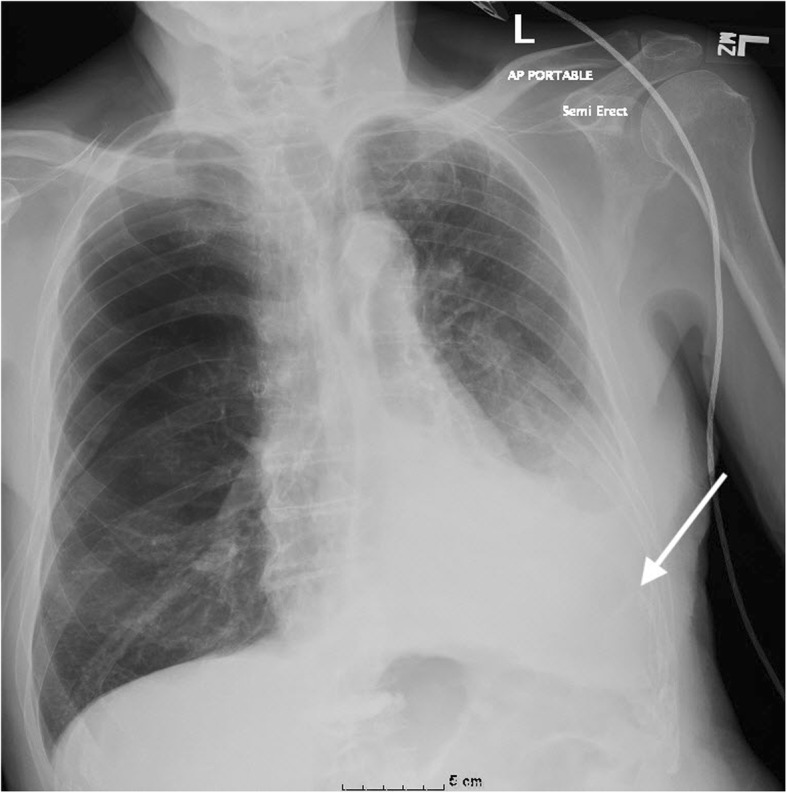
AP (anteroposterior) chest plain radiograph in an 82-year old male, following thoracentesis. Note the significant pleural effusion with compressive atelectasis (arrow) in the left lower hemithorax

AP (anteroposterior) chest plain radiograph in an 82-year old male, following thoracentesis. Note the significant pleural effusion with compressive atelectasis (arrow) in the left lower hemithorax