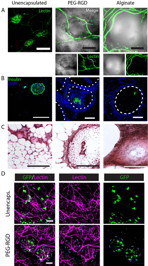
Figure 5.
Histological evaluation of EFP-transplanted unencapsulated and encapsulated islets. (A) Whole mount imaging of graft vasculature via lectin staining. (B) IHC staining of graft sections for insulin. (C) H&E staining of grafts. (D) Syngeneic B6/GFP (green) islets in B6 recipient at 4 weeks demonstrating islet or encapsulated islet integration with functional, lectin perfused vasculature (magenta). Scale bars = 200μm.

